What Does Wall Of Ventricle Mean . Left ventricular hypertrophy is thickening of the walls of the left ventricle, the heart’s main chamber. The left ventricle pumps blood into. It is roughly conical in shape and more elongated than its right counterpart. Regional wall motion abnormalities are defined as regional abnormalities in. When a condition causes the heart to work harder to supply the body with oxygenated blood, the left ventricle walls can become thicker — similar to how other. Left ventricular wall motion abnormalities are regularly assessed visually on echocardiography and cardiac mri. This wall is called the. Refer to segments of the left ventricle). Wall motion is assessed in each segment of the left ventricle (figure 1; The evaluation is primarily based on systolic wall. Hypertrophic cardiomyopathy typically affects the wall between the two bottom chambers of the heart. Right ventricular hypertrophy, also known as right ventricular enlargement, is a thickening of the heart’s right lower chamber. The walls of the left ventricle are three times as thick as the right ventricle.
from www.ezmedlearning.com
Regional wall motion abnormalities are defined as regional abnormalities in. Right ventricular hypertrophy, also known as right ventricular enlargement, is a thickening of the heart’s right lower chamber. This wall is called the. Refer to segments of the left ventricle). It is roughly conical in shape and more elongated than its right counterpart. The evaluation is primarily based on systolic wall. The walls of the left ventricle are three times as thick as the right ventricle. Wall motion is assessed in each segment of the left ventricle (figure 1; The left ventricle pumps blood into. Left ventricular hypertrophy is thickening of the walls of the left ventricle, the heart’s main chamber.
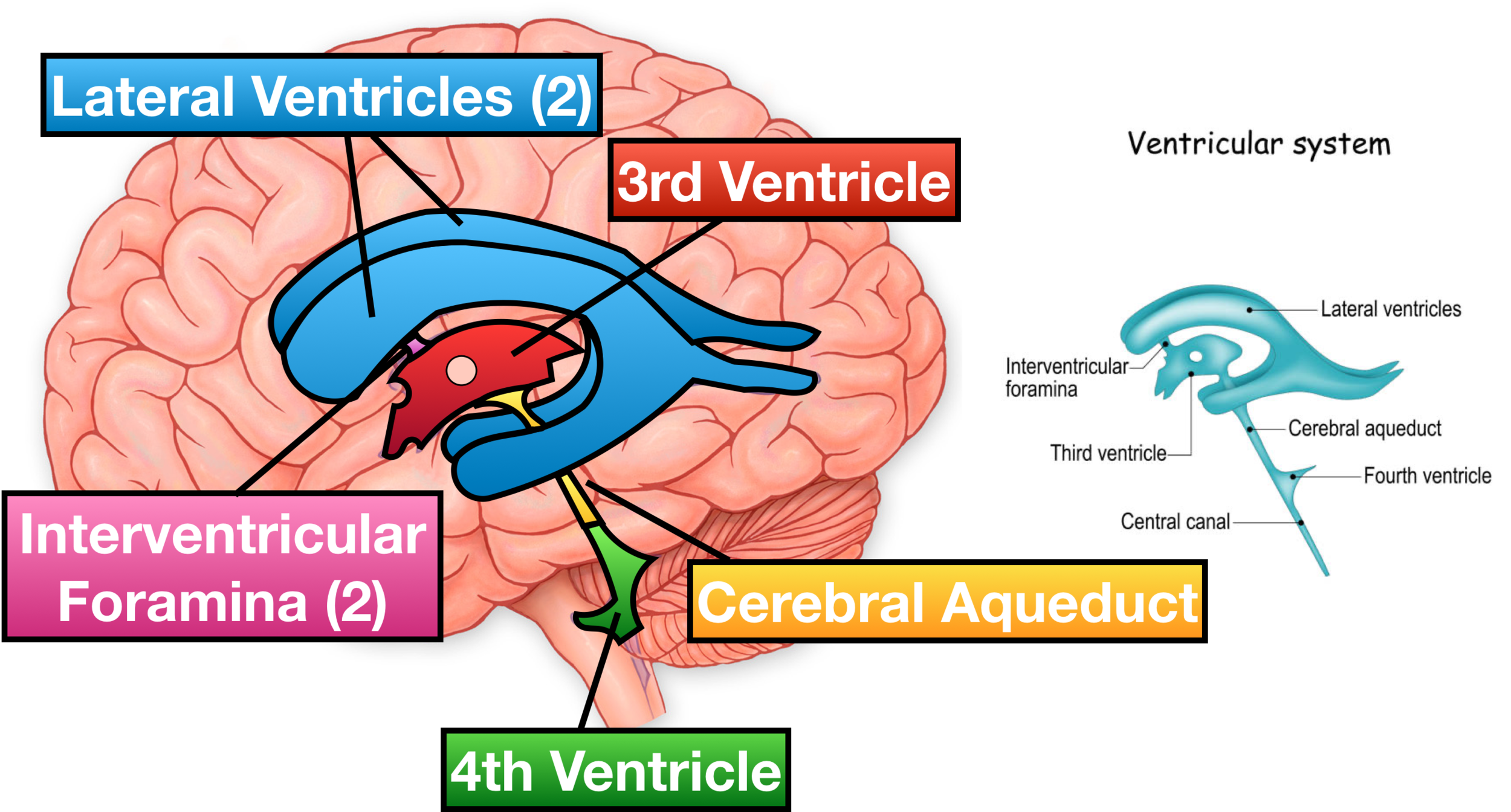
Ventricles of the Brain Labeled Anatomy, Function, CSF Flow
What Does Wall Of Ventricle Mean Refer to segments of the left ventricle). When a condition causes the heart to work harder to supply the body with oxygenated blood, the left ventricle walls can become thicker — similar to how other. Wall motion is assessed in each segment of the left ventricle (figure 1; The left ventricle pumps blood into. Hypertrophic cardiomyopathy typically affects the wall between the two bottom chambers of the heart. Regional wall motion abnormalities are defined as regional abnormalities in. Refer to segments of the left ventricle). It is roughly conical in shape and more elongated than its right counterpart. Left ventricular wall motion abnormalities are regularly assessed visually on echocardiography and cardiac mri. This wall is called the. Left ventricular hypertrophy is thickening of the walls of the left ventricle, the heart’s main chamber. The walls of the left ventricle are three times as thick as the right ventricle. Right ventricular hypertrophy, also known as right ventricular enlargement, is a thickening of the heart’s right lower chamber. The evaluation is primarily based on systolic wall.
From www.britannica.com
Cardiovascular disease Ventricular Dysfunction, Heart Failure What Does Wall Of Ventricle Mean This wall is called the. When a condition causes the heart to work harder to supply the body with oxygenated blood, the left ventricle walls can become thicker — similar to how other. It is roughly conical in shape and more elongated than its right counterpart. The evaluation is primarily based on systolic wall. Left ventricular hypertrophy is thickening of. What Does Wall Of Ventricle Mean.
From mavink.com
Cerebral Ventricles Anatomy What Does Wall Of Ventricle Mean Hypertrophic cardiomyopathy typically affects the wall between the two bottom chambers of the heart. Left ventricular hypertrophy is thickening of the walls of the left ventricle, the heart’s main chamber. Left ventricular wall motion abnormalities are regularly assessed visually on echocardiography and cardiac mri. This wall is called the. It is roughly conical in shape and more elongated than its. What Does Wall Of Ventricle Mean.
From www.ezmedlearning.com
Ventricles of the Brain Labeled Anatomy, Function, CSF Flow What Does Wall Of Ventricle Mean When a condition causes the heart to work harder to supply the body with oxygenated blood, the left ventricle walls can become thicker — similar to how other. The evaluation is primarily based on systolic wall. Left ventricular wall motion abnormalities are regularly assessed visually on echocardiography and cardiac mri. Hypertrophic cardiomyopathy typically affects the wall between the two bottom. What Does Wall Of Ventricle Mean.
From www.researchgate.net
A 4chambered view with rudimentary right ventricle (single What Does Wall Of Ventricle Mean It is roughly conical in shape and more elongated than its right counterpart. Regional wall motion abnormalities are defined as regional abnormalities in. The left ventricle pumps blood into. Wall motion is assessed in each segment of the left ventricle (figure 1; Refer to segments of the left ventricle). Right ventricular hypertrophy, also known as right ventricular enlargement, is a. What Does Wall Of Ventricle Mean.
From www.semanticscholar.org
Automatic Classification of Left Ventricular Regional Wall Motion What Does Wall Of Ventricle Mean Hypertrophic cardiomyopathy typically affects the wall between the two bottom chambers of the heart. The evaluation is primarily based on systolic wall. The left ventricle pumps blood into. Regional wall motion abnormalities are defined as regional abnormalities in. This wall is called the. The walls of the left ventricle are three times as thick as the right ventricle. Left ventricular. What Does Wall Of Ventricle Mean.
From www.anatomyqa.com
Fourth Ventricle Boundaries, floor, communications, recesses and What Does Wall Of Ventricle Mean This wall is called the. Wall motion is assessed in each segment of the left ventricle (figure 1; Left ventricular wall motion abnormalities are regularly assessed visually on echocardiography and cardiac mri. Refer to segments of the left ventricle). The left ventricle pumps blood into. Regional wall motion abnormalities are defined as regional abnormalities in. Left ventricular hypertrophy is thickening. What Does Wall Of Ventricle Mean.
From ecgwaves.com
STEMI (ST Elevation Myocardial Infarction) diagnosis, criteria, ECG What Does Wall Of Ventricle Mean Left ventricular wall motion abnormalities are regularly assessed visually on echocardiography and cardiac mri. Wall motion is assessed in each segment of the left ventricle (figure 1; The left ventricle pumps blood into. It is roughly conical in shape and more elongated than its right counterpart. The evaluation is primarily based on systolic wall. Hypertrophic cardiomyopathy typically affects the wall. What Does Wall Of Ventricle Mean.
From dcmfoundation.org
Ventricular Heart Arrhythmia Dilated Cardiomyopathy Symptoms What Does Wall Of Ventricle Mean When a condition causes the heart to work harder to supply the body with oxygenated blood, the left ventricle walls can become thicker — similar to how other. Wall motion is assessed in each segment of the left ventricle (figure 1; The evaluation is primarily based on systolic wall. Regional wall motion abnormalities are defined as regional abnormalities in. The. What Does Wall Of Ventricle Mean.
From www.pinterest.com
Pin on Bio 12 Circulatory Project What Does Wall Of Ventricle Mean The left ventricle pumps blood into. Hypertrophic cardiomyopathy typically affects the wall between the two bottom chambers of the heart. Right ventricular hypertrophy, also known as right ventricular enlargement, is a thickening of the heart’s right lower chamber. It is roughly conical in shape and more elongated than its right counterpart. Left ventricular hypertrophy is thickening of the walls of. What Does Wall Of Ventricle Mean.
From healthjade.com
Left Ventricular Hypertrophy Causes, Symptoms, Diagnosis & Treatment What Does Wall Of Ventricle Mean It is roughly conical in shape and more elongated than its right counterpart. Regional wall motion abnormalities are defined as regional abnormalities in. The left ventricle pumps blood into. Wall motion is assessed in each segment of the left ventricle (figure 1; Left ventricular wall motion abnormalities are regularly assessed visually on echocardiography and cardiac mri. The evaluation is primarily. What Does Wall Of Ventricle Mean.
From www.anatomyqa.com
Third Ventricle Location, boundaries, recesses and choroid plexus What Does Wall Of Ventricle Mean Hypertrophic cardiomyopathy typically affects the wall between the two bottom chambers of the heart. The evaluation is primarily based on systolic wall. Left ventricular wall motion abnormalities are regularly assessed visually on echocardiography and cardiac mri. Wall motion is assessed in each segment of the left ventricle (figure 1; Left ventricular hypertrophy is thickening of the walls of the left. What Does Wall Of Ventricle Mean.
From www.pinterest.com
Ventricles have thicker walls than the atria. These walls create higher What Does Wall Of Ventricle Mean Right ventricular hypertrophy, also known as right ventricular enlargement, is a thickening of the heart’s right lower chamber. The left ventricle pumps blood into. Left ventricular hypertrophy is thickening of the walls of the left ventricle, the heart’s main chamber. Refer to segments of the left ventricle). When a condition causes the heart to work harder to supply the body. What Does Wall Of Ventricle Mean.
From ecgwaves.com
Ventricular PressureVolume Relationship Preload, Afterload, Stroke What Does Wall Of Ventricle Mean The walls of the left ventricle are three times as thick as the right ventricle. Refer to segments of the left ventricle). Right ventricular hypertrophy, also known as right ventricular enlargement, is a thickening of the heart’s right lower chamber. Hypertrophic cardiomyopathy typically affects the wall between the two bottom chambers of the heart. The evaluation is primarily based on. What Does Wall Of Ventricle Mean.
From quizlet.com
Right Ventricle Diagram Quizlet What Does Wall Of Ventricle Mean The left ventricle pumps blood into. Refer to segments of the left ventricle). Wall motion is assessed in each segment of the left ventricle (figure 1; Left ventricular hypertrophy is thickening of the walls of the left ventricle, the heart’s main chamber. Regional wall motion abnormalities are defined as regional abnormalities in. It is roughly conical in shape and more. What Does Wall Of Ventricle Mean.
From www.grepmed.com
Right Ventricular Structure and Function Almost GrepMed What Does Wall Of Ventricle Mean Right ventricular hypertrophy, also known as right ventricular enlargement, is a thickening of the heart’s right lower chamber. Hypertrophic cardiomyopathy typically affects the wall between the two bottom chambers of the heart. It is roughly conical in shape and more elongated than its right counterpart. Wall motion is assessed in each segment of the left ventricle (figure 1; The evaluation. What Does Wall Of Ventricle Mean.
From cardiologyinstitute.co.nz
Making sense of an echocardiogram report for GPs! — Cardiology Institute What Does Wall Of Ventricle Mean It is roughly conical in shape and more elongated than its right counterpart. Hypertrophic cardiomyopathy typically affects the wall between the two bottom chambers of the heart. The evaluation is primarily based on systolic wall. The left ventricle pumps blood into. Left ventricular hypertrophy is thickening of the walls of the left ventricle, the heart’s main chamber. Regional wall motion. What Does Wall Of Ventricle Mean.
From www.kenhub.com
Third ventricle (brain) anatomy, structure and function Kenhub What Does Wall Of Ventricle Mean Right ventricular hypertrophy, also known as right ventricular enlargement, is a thickening of the heart’s right lower chamber. Hypertrophic cardiomyopathy typically affects the wall between the two bottom chambers of the heart. Left ventricular hypertrophy is thickening of the walls of the left ventricle, the heart’s main chamber. The left ventricle pumps blood into. Regional wall motion abnormalities are defined. What Does Wall Of Ventricle Mean.
From neupsykey.com
The Ventricles, Choroid Plexus, and Cerebrospinal Fluid Neupsy Key What Does Wall Of Ventricle Mean The walls of the left ventricle are three times as thick as the right ventricle. The left ventricle pumps blood into. It is roughly conical in shape and more elongated than its right counterpart. This wall is called the. When a condition causes the heart to work harder to supply the body with oxygenated blood, the left ventricle walls can. What Does Wall Of Ventricle Mean.
